The connection between ligusticum chuanxiong and MIRI
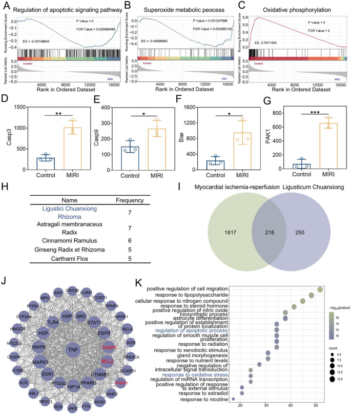
MIRI is among the most extreme problems following surgical operations for myocardial infarction in scientific apply [33]. An in-depth evaluation of the connection between MIRI and oxidative stress, in addition to its particular pathogenesis, is essential for enhancing scientific therapy and enhancing affected person prognosis. To totally discover the pathogenesis of MIRI, this examine obtained high-throughput sequencing expression profile information from the Gene Expression Omnibus (GEO) and performed analysis utilizing the Gene Set Enrichment Evaluation (GSEA) methodology. The gene expression datasets of the hearts from the management group and MIRI-modeled rats (GEO acc + ession quantity: GSE240847) have been utilized on this examine. GSEA outcomes confirmed that DEGs have been considerably enriched in apoptosis-related signaling pathways throughout the Gene Ontology (GO) gene set (Fig. 2A). In the meantime, the analysis findings additionally demonstrated that the pathogenesis of MIRI is carefully linked to a number of redox-related pathways (Fig. 2B, C, and Fig. S1). This examine additional examined a number of genes related to apoptosis, particularly Caspase9, Caspase3, Bax, and PAK1, by analyzing their expression ranges. The outcomes indicated that these genes have been upregulated within the MIRI rat mannequin, confirming the activation of the apoptosis pathway. This discovering establishes a robust correlation between MIRI and apoptosis (Fig. 2D-G).
Ligusticum Chuanxiong has an extended historical past of use in treating coronary heart illnesses.8 In present scientific apply, it’s usually used as an adjuvant drug for the therapy of MIRI. Nonetheless, the intrinsic relationship between Ligusticum Chuanxiong and MIRI stays unclear. This analysis explored the hyperlink between Ligusticum Chuanxiong and MIRI by means of drug screening and community pharmacology. Firstly, by means of drug screening, Ligusticum Chuanxiong was recognized as a key conventional Chinese language drugs for the therapy of MIRI. Utilizing “myocardial ischemia-reperfusion” because the key phrase, related Conventional Chinese language drugs prescriptions have been retrieved from the prescription database of Huabing Knowledge (http://www.huabeing.com), and the medication within the prescriptions have been sorted and analyzed. The outcomes confirmed that Ligusticum Chuanxiong and Astragali membranaceus have been the medication with the very best frequency of use (Fig. 2H). Subsequently, all of the lively substances of Ligusticum Chuanxiong have been mined utilizing the Conventional Chinese language Medication Techniques Pharmacology Database and Evaluation Platform (TCMSP, https://tcmsp-e.com/tcmsp.php). Given the quite a few lively substances of Ligusticum Chuanxiong, screening situations have been set primarily based on pharmacokinetic parameters. Oral bioavailability (OB) ≥ 30% and drug-likeness (DL) ≥ 0.18 have been set because the screening standards. Lastly, 10 lively substances have been chosen because the analysis objects (Desk S1). These 10 lively substances have been uploaded to the Swiss Goal Prediction platform for goal evaluation, yielding 672 potential targets. On the identical time, 2036 targets associated to myocardial ischemia-reperfusion have been obtained from the Gene Playing cards database. The overlap between Ligusticum Chuanxiong’s predicted targets and myocardial ischemia-reperfusion targets was analyzed, leading to 218 shared targets (Fig. 2I). These 218 mapped targets have been uploaded to the STRING database to generate the TSV file of the PPI community, subsequently analyzed utilizing Cytoscape 3.10.1. In Cytoscape, targets with excessive dispersion have been eliminated, and the “Community Analyzer” operate was used to judge the topological traits of the outcomes. The highest 40 targets with the very best diploma values have been chosen as key targets. Visualization was carried out on the 40 key targets obtained by screening to assemble and analyze the protein-protein interplay community (Fig. 2J). Lastly, the 40 key targets have been uploaded to the Metascape database for Gene Ontology (GO) pathway enrichment evaluation. The findings revealed that Ligusticum Chuanxiong’s connection to MIRI is strongly linked to oxidative stress and apoptosis signaling pathways (Fig. 2Ok). In conclusion, we imagine that Ligusticum Chuanxiong is a promising drug for decreasing MIRI by concentrating on oxidative stress and apoptosis pathways.
The connection between Ligusticum Chuanxiong and MIRI. (A–C) GSEA of differentially expressed genes between the Management group and MIRI rats (GEO accession no. 240847). (D–G) Comparability of the gene expression ranges of the apoptosis signaling pathway amongst totally different teams (Casp3, Casp9, Bax, and PAK1). (H) The highest 5 medication when it comes to frequency. (I) Venn diagram of the intersection of the targets of the lively substances of Ligusticum Chuanxiong and disease-related targets. (J) Protein-protein interplay community of 40 key targets. (Ok) GO pathway enrichment evaluation. Statistical comparisons have been made utilizing one-way ANOVA and t-test. Statistical significance was indicated as *p < 0.05, **p < 0.01, ***p < 0.001. Knowledge are expressed as imply ± SD (n = 3)
Synthesis and characterization of LC-CDs
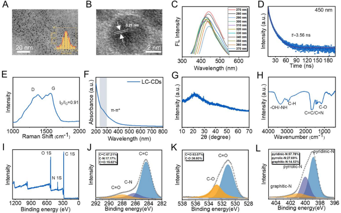
The difficulties in isolating the lively constituents of Ligusticum Chuanxiong, coupled with its restricted oral bioavailability and extra constraints, have impeded its utility in scientific apply. To successfully deal with this problem, LC-CDs have been efficiently synthesized utilizing a easy one-step hydrothermal methodology. The recent Chinese language natural drugs Ligusticum chuanxiong was dried and floor into powder. Subsequently, LC-CDs have been efficiently obtained by means of a easy one-step hydrothermal synthesis methodology. Throughout a number of synthesis processes, the yields of LC-CDs have been comparatively comparable, roughly 10%. The transmission electron microscopy (TEM) picture of LC-CDs is introduced in Fig. 3A. The picture demonstrates that LC-CDs possess wonderful dispersibility and exhibit a morphology that’s roughly quasi-spherical. The inset in Fig. 3A is the statistical results of the particle sizes of LC-CDs. This means that LC-CDs exhibit a slim dimension distribution, with a mean diameter of round 2.4 ± 0.03 nm (Fig. 3A). The extraordinarily small particle dimension of LC-CDs signifies that it has a excessive particular floor space, which allows LC-CDs to reveal extra lively websites and thus totally exert their capabilities. To additional examine the microscopic structural traits of LC-CDs, a high-resolution transmission electron microscope (HRTEM) was used to meticulously study their lattice. After exact measurement, the lattice spacing of LC-CDs was discovered to be 0.21 nm, which corresponds to the (100) crystal aircraft of graphite, strongly demonstrating that LC-CDs possess a graphite-like part construction (Fig. 3B). When CDs possess graphitized lattices with a spacing of 0.21 nm, their atomic association displays a extremely ordered π-π conjugate system. This structural characteristic offers a great situation for the formation of delocalized giant π bonds. Within the catalytic course of, the presence of delocalized giant π bonds considerably enhances the delocalization diploma of the electron cloud, not solely decreasing the electron switch resistance of the system but in addition successfully selling the directional migration of electrons between the lively websites and reactants, thus remarkably enhancing the electron switch effectivity and total efficiency of the catalytic response. The fluorescence property is among the key properties of CDs. This analysis totally investigated the fluorescence conduct of LC-CDs underneath various excitation wavelengths. As proven in Fig. 3C underneath a number of excitation wavelengths, the optimum emission wavelengths of LC-CDs have been predominantly concentrated round 450 nm. Additional, by detecting the fluorescence decay curve of LC-CDs and thru exact calculation, its fluorescence lifetime was obtained as 3.56 ns (Fig. 3D). In a darkish atmosphere, when LC-CDs have been irradiated with ultraviolet gentle, it could possibly be noticed that they emitted blue-green fluorescence (Fig. S2). The above-mentioned phenomena totally point out that the CDs derived from Ligusticum Chuanxiong have been efficiently synthesized. Raman spectroscopy was used to precisely measure the diploma of graphitization and floor defects in LC-CDs. Within the Raman spectrum, the D peak round 1300 cm⁻¹ displays the defect state of the carbon lattice, whereas the G peak close to 1580 cm⁻¹ corresponds to the sp² hybridized carbon’s in-plane vibrational mode, indicating the graphitization degree of LC-CDs. Raman spectroscopy revealed the coexistence of D and G peaks within the LC-CDs spectrum, with an ID/IG ratio of 0.91 (Fig. 3E). This outcome signifies that LC-CDs not solely have a comparatively excessive graphitization diploma but in addition have a lot of defect websites on their floor. These floor defects and the π-π planar construction conducive to speedy electron switch are extremely prone to endow LC-CDs with outstanding catalytic properties. The ultraviolet absorption spectroscopy evaluation outcomes confirmed that LC-CDs exhibited a comparatively broad absorption band within the wavelength vary of 200–600 nm (Fig. 3F). This phenomenon is especially attributed to the π-π* transition and n-π* transition present within the carbon-dot construction. Amongst them, the π-π* transition originates from the presence of the sp²-hybridized construction within the carbon core. Beneath ultraviolet gentle irradiation, π-bonded electrons are excited from the π orbital to the π* orbital [34]. The n-π transition arises from floor purposeful teams like -NH₂, -OH, and -COOH, enabling non-bonding electrons to transition to the π orbital upon photon absorption. These purposeful teams additionally present a cloth foundation for LC-CDs to exert their capabilities. The X-ray diffraction (XRD) experiment additional characterised the crystal construction of LC-CDs. The XRD sample confirmed that LC-CDs have a comparatively broad diffraction peak at 21.4°, which could possibly be attributed to the (002) aircraft of graphite. Via calculation, the d-spacing similar to this diffraction peak was roughly 0.42 nm (Fig. 3G). Subsequently, Fourier-transform infrared spectroscopy (FT-IR) and X-ray photoelectron spectroscopy (XPS) have been used to additional and meticulously characterize the floor construction of LC-CDs. Within the FT-IR spectrum, the broad absorption peak at 3375 cm⁻¹ could possibly be attributed to the stretching vibration of-OH/-NH; the absorption peak at 2950 cm⁻¹ originated from the symmetric and uneven stretching vibrations of methyl and methylene teams (C-H) in saturated hydrocarbons; the absorption peak at 1661 cm⁻¹ corresponded to the absorption peak of the C = C double bond within the conjugated system or the stretching vibration of the C = N bond; the absorption peak at 1180 cm⁻¹ was brought on by the stretching vibration of the C-O bond (Fig. 3H). These peaks verify that LC-CDs possess ample floor purposeful teams. The XPS evaluation confirmed that LC-CDs are primarily composed of three parts: C, N, and O. Amongst them, the C1s peak had the very best depth, with a corresponding content material of 73.01%; the contents of O1s and N1s are 19.87% and seven.12%, respectively (Fig. 3I). Additional, the XPS spectrum was subjected to peak-fitting processing for various parts. Within the C1s spectrum, the height at 248.4 eV corresponded to the C = C bond, the height at 286.3 eV corresponded to the C-N bond, and the height at 287.9 eV corresponded to the C = O; within the O1s spectrum, the height at 531.1 eV corresponded to the C = O, and the height at 532.8 eV corresponded to the C-O; within the N1s spectrum, the peaks at 398.9 eV, 400.07 eV, and 400.93 eV represented pyridinic-N, pyrrolic-N, and graphitic-N respectively. The detailed evaluation outcomes confirmed that within the C1s spectrum, the content material of C = C was the very best, reaching 67.21%, and the contents of C-N and C = O have been 17.17% and 15.62% respectively; within the O1s spectrum, the contents of C = O and C-O have been 63.07% and 36.93% respectively; within the N1s spectrum, the contents of pyridinic-N, pyrrolic-N, and graphitic-N have been 57.78%, 27.69%, and 14.53% respectively (Fig. 3J-L). The characterization outcomes of XPS conclusively verify that the chemical teams contained in Ligusticum chuanxiong are retained on the floor of LC-CDs. These teams represent the fabric foundation for LC-CDs to exert their pharmacological actions, offering essential structural help for the belief of their organic capabilities. In conclusion, our thorough and systematic structural characterization of LC-CDs has confirmed their profitable synthesis. The abundance of floor purposeful teams on LC-CDs allows potential pharmacological and catalytic functionalities. This discovering tremendously motivates us to additional discover the potential functions of LC-CDs in related fields.
Synthesis and characterization of LC-CDs. (A) TEM and dimension distribution histogram of LC-CDs. (B) HR-TEM of Ca-CDs. (C) Emission spectra of LC-CDs underneath totally different excitations. (D) Fluorescence lifetime of LC-CDs. (E) Raman spectrum and (F) UV-vis spectrum of LC-CDs. (G) XRD spectrum and (H) FT-IR spectrum of LC-CDs. (I) XPS survey spectra of LC-CDs. (J–L) Excessive-resolution XPS spectra of C 1s, O 1s, and N 1s for LC-CDs
Free radical scavenging analysis of LC-CDs
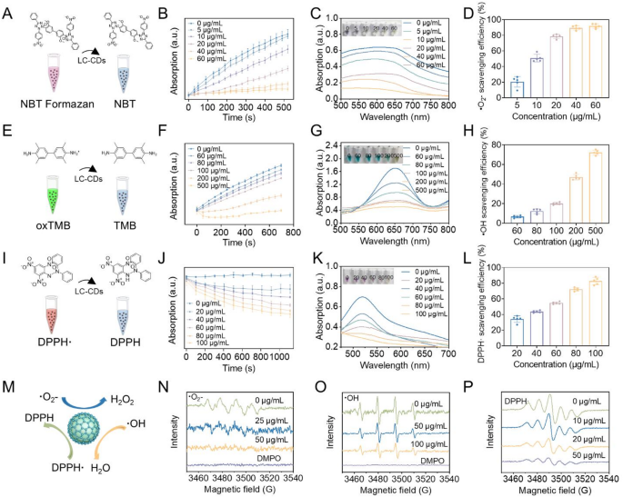
Inside the carbon core construction, carbon atoms are organized in a carefully packed method [35]. This extremely ordered association sample can considerably cut back the power hole width between carbon atoms [36]. In accordance with the band idea, the discount of the power hole width implies a lower within the power required for electron transition [36]. Valence band electrons will be readily promoted by means of the absorption of modest power inputs to traverse the forbidden power hole and populate the conduction band. This digital transition mechanism facilitates a marked enhance in cost provider density, because the delocalized electrons within the conduction band exhibit considerably enhanced mobility. The resultant elevation in free provider inhabitants immediately correlates with improved cost transport traits, thereby considerably enhancing the carbon-core materials’s conductivity efficiency. The superb electron-transfer properties of CDs can endow them with good catalytic exercise. The analysis by Gao et al. has demonstrated that CDs derived from activated carbon possess ultra-high SOD exercise [26]. Via surface-group capping experiments and quite a few research, it has been proven that the SOD exercise of CDs stems from the mix of hydroxyl and carboxyl teams with superoxide anions, in addition to the electron switch carried out by carbonyl teams conjugated with the π-system. In our earlier analysis, we additionally verified that CDs derived from Honeysuckle (Hy-CDs) exhibit robust SOD exercise. In that analysis, we verified that the SOD exercise of Hy-CDs is mainly derived from a considerable amount of amino teams on their floor. Constructing on this basis, we systematically investigated the catalytic properties of LC-CDs. First, we examined the flexibility of LC-CDs to scavenge ·O₂⁻. A technology system of ·O₂⁻ was constructed utilizing riboflavin and methionine. Upon gentle publicity, riboflavin absorbs photon power, undergoes excitation, and donates electrons to methionine, self-oxidizing to generate a semiquinone radical. The semiquinone radical reacts with molecular oxygen to generate ·O₂⁻. The ·O₂⁻ generated within the system oxidizes nitroblue tetrazolium chloride (NBT) to blue-violet NBT formazan, which has a robust absorption at 560 nm (Fig. 4A). This method can be utilized to detect the scavenging impact of LC-CDs on ·O₂⁻. We first examined the kinetics of LC-CDs in scavenging ·O₂⁻ utilizing this technique. The UV absorption of options containing totally different concentrations of LC-CDs at 560 nm was constantly monitored inside 520s. The outcomes confirmed that as time elapsed, the absorption of the system answer with out LC-CDs at 560 nm step by step elevated, indicating that a considerable amount of ·O₂⁻ was generated within the answer. Nonetheless, after including LC-CDs, the UV absorption of the answer decreased considerably, suggesting that LC-CDs can remarkably inhibit the technology of ·O₂⁻ (Fig. 4B). Particularly because the focus of LC-CDs attained 60 µg/mL, the absorption of the answer at 560 nm hardly modified. Subsequent UV-Vis spectral evaluation (500–800 nm) of the post-reaction answer revealed absorption traits that carefully mirrored the time-dependent kinetic conduct noticed within the chemical response (Fig. 4C). The ultimate statistical outcomes confirmed that because the focus of LC-CDs elevated, their capability to scavenge ·O₂⁻ step by step enhanced. Because the focus of LC-CDs attained 60 µg/mL, the scavenging charge of ·O₂⁻ practically reached 100% (Fig. 4D). These outcomes point out that LC-CDs possess robust SOD-like enzyme exercise. Instantly after, we examined the flexibility of LC-CDs to scavenge hydroxyl radicals (·OH). ·OH is a key factor inflicting and aggravating the physiological imbalance within the human physique. We simulated the technology of ·OH utilizing the Fenton response between Fe²⁺ and H₂O₂. On this system, 3,3’,5,5’-tetramethylbenzidine (TMB) was used as a chromogenic substance (Fig. 4E). After being oxidized to oxTMB, the answer turns inexperienced and will be detected at 652 nm by a microplate reader. Constant kinetic detection outcomes indicated that with the progressive enhance within the focus of LC-CDs, the oxidation charge of TMB within the answer exhibited a gradual deceleration. This discovering corroborated the truth that LC-CDs possess a particular capability for scavenging·OH (Fig. 4F). Additional, we detected the UV absorption spectrum of response techniques with totally different LC-CDs concentrations within the vary of 500–800 nm. The info revealed that when the focus of LC-CDs reached 500 µg/mL, TMB was hardly oxidized (Fig. 4G). The statistical outcomes confirmed that high-concentration LC-CDs can obtain a scavenging charge of practically 80% for ·OH (Fig. 4H).
Thereafter, we evaluated the scavenging impact of LC-CDs on nitrogen-containing radicals. 1,1-Diphenyl-2-picrylhydrazyl radical (DPPH·) is a typical nitrogen-containing radical. The lone-pair electrons of DPPH· have robust absorption within the seen gentle area (515–520 nm), making the answer purple (Fig. 4I). The kinetic outcomes confirmed that the scavenging impact of LC-CDs on DPPH· is concentration-dependent. As time elevated, LC-CDs at totally different concentrations all demonstrated scavenging effectivity for DPPH· (Fig. 4J). The UV absorption spectrum outcomes confirmed that because the focus of LC-CDs reached 100 µg/mL, the answer nearly pale, indicating that DPPH· within the answer had been utterly scavenged (Fig. 4Ok). The statistical outcomes additionally confirmed that because the focus of LC-CDs reached 100 µg/mL, the scavenging charge for DPPH· might attain roughly 80% (Fig. 4L). Subsequent, we used electron paramagnetic resonance (EPR) know-how to additional detect the scavenging impact of LC-CDs on ·O₂⁻, ·OH, and DPPH· (Fig. 4N-P). The qualitative outcomes indicated that LC-CDs have an excellent scavenging impact on these three radicals, and it’s concentration-dependent. All of the above-mentioned outcomes recommend that LC-CDs possess robust antioxidant capability (Fig. 4M). Given the standard pathological characteristic of ROS burst throughout myocardial ischemia-reperfusion, the antioxidant properties of LC-CDs are anticipated to point out potential utility worth within the prophylaxis and administration methods for this illness.
Free radical scavenging analysis of LC-CDs. (A) Schematic diagram of superoxide radical scavenging system. (B) Kinetic curves of ·O₂⁻ scavenged by LC-CDs. (C) UV absorption spectra of ·O₂⁻ scavenged by LC-CDs. (D) The scavenging charge of ·O₂⁻ by LC-CDs. (E) Schematic diagram of ·OH scavenging system. (F) Kinetic curves of ·OH scavenged by LC-CDs. (G) UV absorption spectra of ·OH scavenged by LC-CDs. (H) The scavenging charge of ·OH by LC-CDs. (I) Schematic diagram of DPPH· scavenging system. (J) Kinetic curves of DPPH· scavenged by LC-CDs. (Ok) UV absorption spectra of DPPH· scavenged by LC-CDs. (L) The scavenging charge of DPPH· by LC-CDs. (M) Schematic diagram of the scavenging of three kinds of free radicals by LC-CDs. (N–P) EPR Spectra Detect the Scavenging of ·O₂⁻, ·OH, and DPPH· by LC-CDs. Knowledge are expressed as imply ± SD (n = 3)
Sub-organelle co-localization and antioxidant properties of LC-CDs in cells
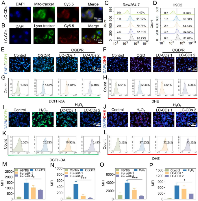
Upon myocardial ischemia-reperfusion, the restoration of nutrient and oxygen provide, nonetheless, exacerbates the technology of ROS [37]. The instantaneous burst of ROS disrupts the redox homeostasis within the native cardiac area, thereby inflicting injury to myocardial cells and additional inducing inflammatory responses [38]. On this course of, the interplay between irritation and ROS collectively aggravates the MIRI. To totally examine the restore efficacy of LC-CDs on MIRI, this examine systematically examined the antioxidant and anti inflammatory capacities of LC-CDs on the mobile degree. Firstly, the generally used immune cell mannequin, Raw264.7 cells, was chosen to discover the localization of LC-CDs inside subcellular organelles. The burst of ROS normally originates from mitochondria. Within the mitochondrial respiratory chain, when electrons fail to switch easily to coenzyme Q, they may leak out and immediately react with molecular oxygen, producing a considerable amount of ·O₂⁻, in the end resulting in redox imbalance [39]. Due to this, this examine first evaluated the colocalization of LC-CDs with mitochondria. With a view to obtain direct visualization of liquid-phase LC-CDs utilizing fluorescence microscopy, this examine employed a chemical modification methodology to connect the fluorescent dye Cy5.5-amino to the floor of LC-CDs. After incubation therapy, the fluorescence emitted by mitochondria and LC-CDs could possibly be clearly noticed underneath a confocal laser scanning microscope. Via the picture merging operation, a lot of overlapping areas between them have been introduced (Fig. 5A). Fluorescence colocalization evaluation confirmed that the Pearson correlation coefficient of colocalization between mitochondria and LC-CDs was as excessive as 0.98 (Fig. S3A, B). This outcome totally confirms that LC-CDs are extremely localized in mitochondria, and this localization attribute facilitates their environment friendly scavenging of ROS. Subsequently, we evaluated the colocalization of LC-CDs with lysosomes. The outcomes indicated that LC-CDs exhibited a sure diploma of colocalization with lysosomes, however in contrast with mitochondria, the colocalization impact was comparatively weaker (Fig. 5B). Via evaluation, the colocalization coefficient of LC-CDs with lysosomes was 0.79 (Fig. S3C, D). The above outcomes recommend that LC-CDs usually tend to localize in mitochondria and comparatively simple to flee from lysosomes. Subsequently, this examine employed stream cytometry to judge the uptake of LC-CDs by Raw264.7 and H9C2 cells. It was discovered that after 1-hour incubation, the uptake charge of LC-CDs by Raw264.7 cells might attain 64.10%, and that by H9C2 cells might attain 36.80% (Fig. 5C, D). Because the incubation time extended, the uptake of LC-CDs by each cell sorts elevated considerably. Subsequent, an OGD/R mannequin was constructed on the mobile degree on this examine to simulate the oxidative stress induced by ischemia-reperfusion. The two’,7’-dichlorodihydrofluorescein (DCFH) probe was utilized to observe the intracellular ROS degree. When the intracellular oxygen strain elevated, DCFH was oxidized to DCF, which emits robust inexperienced fluorescence. The outcomes confirmed that oxidative stress was efficiently induced in H9C2 cells underneath OGD/R therapy. Nonetheless, after therapy with LC-CDs, the fluorescence depth of DCF was considerably weakened (Fig. 5E). In the meantime, dihydroethidium (DHE) was utilized to particularly assess the focus of ·O₂⁻ in cells, and the outcomes demonstrated that LC-CDs might additionally considerably scavenge ·O₂⁻ induced by OGD/R (Fig. 5F). The stream cytometry outcomes strongly confirmed that LC-CDs possess outstanding antioxidant capability within the OGD/R mannequin (Fig. 5G, H). Moreover, this examine used H₂O₂ to induce Raw264.7 cells to assemble a secondary oxidative stress cell mannequin. Equally, DCFH and DHE have been used to detect the degrees of ROS and ·O₂⁻ in Raw264.7 cells. The findings demonstrated that in therapy with LC-CDs, the degrees of ROS and ·O₂⁻ in Raw264.7 cells decreased considerably (Fig. 5I, J). The outcomes from stream cytometry additional quantified the flexibility of LC-CDs to scavenge ROS inside cells. The stream cytometry outcomes confirmed that LC-CDs might even cut back the extent of ·O₂⁻ to that of the management group (Fig. 5Ok, L). Equally, the outcomes obtained from fluorescence statistics of fluorescent pictures additionally help the above conclusion (Fig. 5M-P). The above collection of outcomes totally verify that LC-CDs additionally possess robust antioxidant capability on the mobile degree.
Sub-organelle co-localization and antioxidant properties of LC-CDs in cells. (A) Fluorescence colocalization of LC-CDs with mitochondria. (B) Fluorescence colocalization of LC-CDs with lysosomes. (C and D) Circulation cytometric evaluation of the uptake of LC-CDs by Raw264.7 cells and H9C2 cells at totally different occasions. (E and F) Fluorescence pictures of various teams of H9C2 cells incubated with DCFH and DHE. (G and H) Circulation evaluation outcomes of various teams of H9C2 cells incubated with DCFH and DHE. (I and J) Fluorescence pictures of various teams of Raw264.7 cells incubated with DCFH and DHE. (Ok and L) Circulation evaluation outcomes of various teams of Raw264.7 cells incubated with DCFH and DHE. (M and N) MFI statistics after incubation of DCFH and DHE with H9C2 cells in several teams. (O and P) MFI statistics after incubation of DCFH and DHE with Raw264.7 cells in several teams. LC-CDs 1: inducer + 5 µg/mL LCCDs; LC-CDs 2: inducer + 10 µg/mL LC-CDs. Statistical comparisons have been made utilizing one-way ANOVA and a t-test. Statistical significance was indicated as *p < 0.05, **p < 0.01, ***p < 0.001. Knowledge are expressed as imply ± SD (n = 3)
Cytoprotective impact of LC-CDs
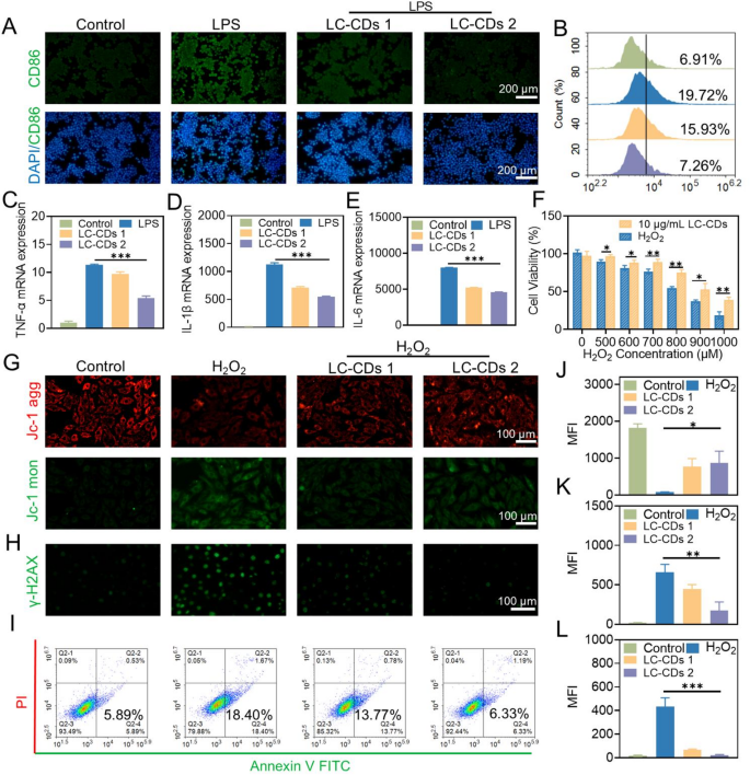
A number of research have demonstrated that macrophages are susceptible to polarize into M1-type macrophages underneath the stimulation of ROS. M1-type macrophages secrete a considerable amount of pro-inflammatory cytokines, which exacerbate the diploma of harm following myocardial ischemia-reperfusion [40,41,42]. On this examine, lipopolysaccharide (LPS) was employed to induce Raw264.7 cells to assemble an M1-type macrophage mannequin, and the cell-surface marker CD86 was chosen to determine M1-type macrophages. The outcomes of immunofluorescence detection confirmed that after therapy with LPS, LC-CDs at totally different concentrations might considerably inhibit the over-expression of CD86 on the floor of macrophages, and this outcome was additionally confirmed by stream cytometry experiments (Fig. 6A, B). Furthermore, the massive variety of inflammatory cytokines secreted by M1-type macrophages aggravates the pathological strategy of myocardial ischemia-reperfusion. Based mostly on this, this examine evaluated the impact of LC-CDs on the discharge of pro-inflammatory cytokines (reminiscent of TNF-α, IL-1β, and IL-6) by Raw264.7 cells on the gene-expression degree. The outcomes of real-time fluorescence qPCR indicated that, underneath the stimulation of LPS, the expression of TNF-α, IL-1β, and IL-6 in Raw264.7 cells was considerably upregulated; whereas subsequent therapy with various concentrations of LC-CDs resulted in a dose-dependent suppression of those inflammatory markers (Fig. 6C-E). This totally demonstrates that LC-CDs can considerably inhibit the incidence of the immune response of Raw264.7 cells underneath stimulated situations. The oxidative injury brought on by myocardial ischemia-reperfusion is normally irreversible. Provided that the antioxidant and anti inflammatory results of LC-CDs have been evaluated beforehand, it’s hypothesized that LC-CDs possess a sure capability to withstand oxidative injury. The totally different concentrations of H₂O₂ have been used to induce oxidative stress in cells, thereby triggering cell loss of life. The outcomes of the 3-(4,5-dimethylthiazol-2-yl)-2,5-diphenyltetrazolium bromide (MTT) assay confirmed that LC-CDs at a focus of 10 µg/mL might successfully reverse the cell loss of life induced by H₂O₂ (Fig. 6F). Throughout ischemia-reperfusion damage, ROS overproduction targets unsaturated fatty acids throughout the mitochondrial membrane, inflicting structural and purposeful injury that impairs ion and metabolite transport [43]. On this examine, the JC-1 fluorescent probe was employed to judge modifications in mitochondrial membrane potential in H9C2 cells following H₂O₂ stimulation. It was discovered that after H₂O₂ stimulation, the JC-1 aggregates decreased considerably, and the JC-1 monomers elevated, indicating that oxidative stress broken the mitochondrial membrane and severely disrupted mitochondrial operate (Fig. 6G). Nonetheless, after therapy with LC-CDs at totally different concentrations, the mitochondrial membrane potential underwent a notable restoration, and the outcomes of fluorescence statistical evaluation additionally supported this conclusion (Fig. 6J, Ok). Throughout ischemia-reperfusion damage, a discount in mitochondrial membrane potential triggers cytochrome C translocation into the cytoplasm, resulting in the activation of apoptosis-related proteins. LC-CDs defend the energy-metabolism core construction of myocardial cells and successfully forestall the incidence of apoptosis by sustaining the mitochondrial membrane potential underneath oxidative stress. After myocardial ischemia-reperfusion, the extraordinary oxidative stress imbalance and inflammatory response can result in extreme DNA injury [44]. Particularly, ROS can react with DNA bases and deoxyribose, inflicting injury to bases, glycosyl teams, and chains, in the end resulting in extreme cell apoptosis and myocardial fibrosis. Immunofluorescence staining was used to detect the protecting impact of LC-CDs on DNA injury induced by H₂O₂, with γ-H2AX as a marker of DNA injury. The findings revealed that LC-CDs at totally different concentrations suppressed γ-H2AX expression, suggesting their capability to mitigate oxidative stress-induced DNA injury in cells (Fig. 6H, L). As well as, the Annexin V/propidium iodide (PI) staining methodology was utilized to determine early and late apoptosis of cells. Throughout early apoptosis, cell membrane integrity is compromised, resulting in the translocation of phosphatidylserine from the interior to the outer membrane floor. Annexin V can bind to phosphatidylserine (PS) on the outer facet of the cell membrane, enabling the identification of early-apoptotic cells by stream cytometry. Circulation cytometry evaluation revealed that LC-CDs successfully suppressed H₂O₂-induced early apoptosis. Because the focus of LC-CDs attained 10 µg/mL, it might nearly utterly inhibit the early apoptosis of cells (Fig. 6I). In conclusion, the above-mentioned analysis outcomes point out that LC-CDs have a protecting impact in opposition to mitochondrial injury, DNA injury, and cell apoptosis brought on by oxidative stress. These results are essential in the course of the strategy of MIRI, implying that LC-CDs might have a promising utility prospect within the remedy for MIRI.
Analysis of the cytoprotective impact of LC-CDs. (A) Fluorescent pictures of Raw264.7 cells labeled with CD86 in several therapy teams. (B) Circulation cytometry outcomes of Raw264.7 cells labeled with CD86 in several therapy teams. (C–E) qPCR evaluation of pro-inflammatory cytokines (TNF-α, IL-1β, and IL-6) in several therapy teams. (F) Cell viability of various therapy teams. (G) Fluorescent pictures of JC-1 probe staining in several therapy teams (JC-1 agg: purple; JC-1 mon: inexperienced). (H) Fluorescent pictures of γ-H2AX labeling in several therapy teams. (I) Circulation cytometry outcomes of Raw264.7 cells stained with Annexin V FITC/PI in several therapy teams. (J and Ok) MFI statistics of JC-1 probe staining in several therapy teams. (L) MFI statistics of γ-H2AX labeling in several therapy teams. LC-CDs 1: inducer + 5 µg/mL LC-CDs; LC-CDs 2: inducer + 10 µg/mL LC-CDs. Statistical comparisons have been made utilizing one-way ANOVA and a t-test. Statistical significance was indicated as *p < 0.05, **p < 0.01, ***p < 0.001. Knowledge are expressed as imply ± SD (n = 3)
The therapeutic impact of LC-CDs on the MIRI mannequin
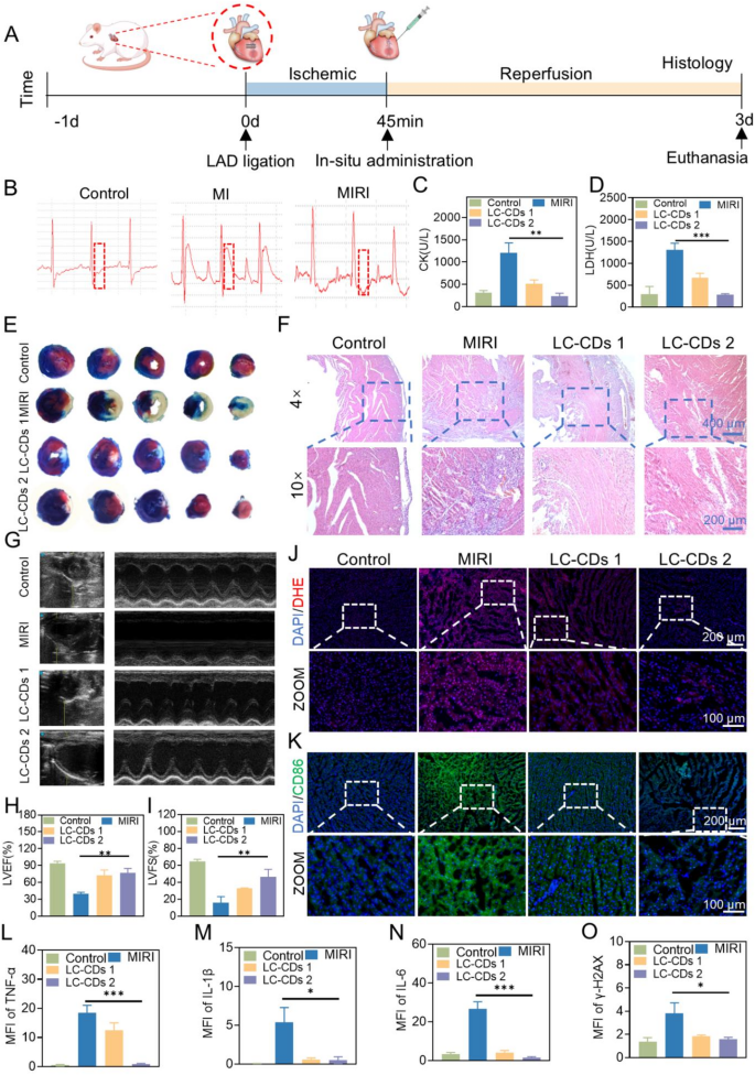
Given the outstanding anti-oxidative injury and anti inflammatory results of LC-CDs on the mobile degree, this investigation arrange a rat mannequin of myocardial ischemia-reperfusion to evaluate the therapeutic efficacy of LC-CDs on the animal degree. The mannequin was constructed by means of surgical procedure (Fig. 7A). Particularly, blood stream within the left anterior descending coronary artery was occluded, adopted by restoration after 45 min to simulate the myocardial ischemia-reperfusion course of. In the meantime, in situ administration was carried out on the rat coronary heart. Three days after administration, the rats underwent transthoracic echocardiography and have been then euthanized. To make sure the soundness of the mannequin, the electrocardiogram (ECG) of the rats was constantly monitored in the course of the examine. Through the myocardial ischemia part, the membrane potential of the ischemic myocardial cells decreases, creating a possible distinction from the conventional myocardium. This led to the technology of an damage present, inflicting the ST-segment elevation within the electrocardiogram leads. Subsequently, ST-segment elevation is essentially the most outstanding electrocardiogram characteristic of myocardial ischemia. Through the 45-minute myocardial ischemia interval, the ST section of the rat coronary heart electrocardiogram was considerably elevated (Fig. 7B). The moment the blood stream was restored, the ST section decreased, which totally demonstrated the profitable development of the rat myocardial ischemia-reperfusion mannequin. Within the therapy part, this examine chosen two concentrations of LC-CDs (1 mg/kg and 5 mg/kg) to deal with the rats to judge their efficacy. LC-CDs have been administered to rats by way of native myocardial injection. When myocardial damage happens, the cell membrane permeability will increase, and creatine kinase (CK) and lactate dehydrogenase (LDH) are launched into the blood. The degrees of CK and LDH within the blood are essential indicators for evaluating cardiac operate. Based mostly on this, the examine collected the serum of handled rats on the similar time level for blood biochemical evaluation. The outcomes confirmed that myocardial damage led to a marked elevation within the ranges of CK and LDH within the blood of the MIRI group rats. After therapy with totally different concentrations of LC-CDs, these two indicators considerably decreased, indicating that the cardiac operate of the handled rats was improved (Fig. 7C, D). To research the infarct space of the rat coronary heart after therapy, this examine employed the TTC (2,3,5-triphenyltetrazolium chloride) and Evans blue staining strategies. TTC will be decreased to purple triphenylformazan (TPF) by dehydrogenases in dwelling tissues, whereas useless cells can not cut back TTC and thus seem white. Evans blue can stain the usually perfused areas of the rat coronary heart tissue blue, and the non-stained areas are the at-risk areas of blood provide. Within the at-risk areas, the elements stained white by TTC are the infarct areas. The staining outcomes of various sections of the rat coronary heart within the MIRI group confirmed that the rat coronary heart had extreme infarction, and nearly all the at-risk areas have been infarcted (Fig. 7E). After therapy with LC-CDs, the infarct space within the at-risk areas decreased. After therapy with a excessive focus of LC-CDs, the infarct space within the at-risk areas nearly returned to regular (Fig. 7E). Given the excellent therapeutic efficacy of LC-CDs, this examine employed paraffin-embedded tissue sectioning and hematoxylin-eosin (H&E) staining to evaluate inflammatory cell infiltration in rat cardiac tissue. The outcomes confirmed that within the infarct space of the MIRI group, a lot of inflammatory cell infiltrations and tissue injury have been seen (Fig. 7F). This injury to the myocardial microenvironment was irreversible. After therapy with totally different concentrations of LC-CDs, the inflammatory infiltration within the infarct space was considerably decreased. In the meantime, LC-CDs additionally decreased the blood cell leakage brought on by ischemia-reperfusion. To guage the influence of LC-CDs on the physiological operate of the rat coronary heart, earlier than euthanizing the rats, transthoracic echocardiography was used to measure the cardiac operate parameters of rats in several teams, together with left ventricular ejection fraction (LVEF), left ventricular fractional shortening (LVFS) (Fig. 7G). As depicted in Fig. 7H, compared with the wholesome group (LVEF: 93.45% ± 4.29), the echocardiogram of the MIRI group rats confirmed weakened left ventricular anterior wall movement (LVEF: 39.99% ± 2.5%), indicating that the myocardial damage brought on by myocardial ischemia-reperfusion would possibly result in cardiac systolic dysfunction. Thankfully, after in situ injection of LC-CDs, the left ventricular anterior wall movement of the rat coronary heart was considerably restored. The LVFS of the low-concentration therapy group reached 72.22% ± 10%, and that of the high-concentration therapy group was 76.62% ± 8.41%. On the identical time, the LVFS of the MIRI group considerably decreased (16.01% ± 7.15%). After therapy with LC-CDs, the LVFS additionally considerably recovered, and the high-concentration therapy group might attain 46.62% ± 8.60% (Fig. 7I). These outcomes totally demonstrated that LC-CDs might restore the myocardial damage brought on by myocardial ischemia and thus restore cardiac operate. The oxidative stress state within the native space of the guts was additional verified by the DHE probe. After ischemia-reperfusion, an unlimited quantity of ROS bursts occurred on the broken website of the guts. After therapy with LC-CDs, the ROS degree was considerably decreased, tremendously assuaging the oxidative injury to myocardial cells (Fig. 7J). Oxidative injury is commonly accompanied by immune infiltration. The earlier H&E staining outcomes have confirmed that LC-CDs can inhibit immune cell infiltration. Additional, by labeling the floor marker CD86 of M1-type macrophages, the modifications in M1-type macrophages within the infarct space of the guts have been noticed. The info indicated a notable rise in CD86 expression within the MIRI group, whereas LC-CDs therapy led to its discount, suggesting a decline in M1-type macrophage ranges throughout the cardiac tissue following LC-CDs administration (Fig. 7Ok). The stream cytometry outcomes of M1 and M2 labeling of myocardial cells in several therapy teams present that after therapy with LC-CDs, it can’t solely cut back the M1 polarization of macrophages but in addition promote the M2 polarization of macrophages (Fig. S4). As well as, immunofluorescence was used to detect the expression ranges of pro-inflammatory cytokines (TNF-α, IL-1β, and IL-6) within the coronary heart tissue after totally different remedies. The findings revealed elevated expression ranges of those three pro-inflammatory cytokines within the MIRI group, with various levels of upregulation. In distinction, the LC-CDs therapy group markedly decreased the degrees of those pro-inflammatory cytokines, contributing to the stabilization of the cardiac immune microenvironment (Fig. S5B-D). The fluorescence statistical information additionally supported this conclusion (Fig. 7L-N). The qPCR outcomes of the myocardial tissues additionally demonstrated the anti-inflammatory effectiveness of LC-CDs (Fig. S6). In the meantime, immunofluorescence of γ-H2AX was additionally used to detect the DNA injury of myocardial cells (Fig. S5A). As anticipated, within the myocardial infarct space, the myocardial cells within the MIRI group had in depth DNA injury, whereas LC-CDs considerably inhibited this injury (Fig. 7O). In conclusion, LC-CDs markedly alleviate cardiac dysfunction induced by myocardial ischemia and defend myocardial cells in opposition to oxidative stress-induced damage. On the identical time, it could actually inhibit immune cell infiltration, decrease the synthesis of pro-inflammatory cytokines, and in the end obtain speedy restore after myocardial ischemia-reperfusion.
The therapeutic impact of LC-CDs on MIRI. (A) Schematic diagram of the experimental process. (B) Electrocardiogram in the course of the institution of rat MIRI mode. (C and D) Contents of CK and LDH within the blood of rats in several therapy teams. (E) Evans blue/TTC staining of coronary heart sections from rats in several therapy teams. (F) H&E pathological part staining of the hearts of rats in several therapy teams. (G) Transthoracic echocardiography of rats in several therapy teams. (H and I) LVEF and LVFS of the hearts of rats in several therapy teams. (J and Ok) DHE and CD86 immunofluorescence staining of the hearts of rats in several therapy teams. (L–O) Statistics of fluorescence depth of immunofluorescence staining (TNF-α, IL-1β, IL-6, and γ-H2AX). LC-CDs 1: MIRI + 1 mg/kg LC-CDs; LC-CDs 2: MIRI + 5 mg/kg LC-CDs. Statistical comparisons have been made utilizing one-way ANOVA and t-test. Statistical significance was indicated as *p < 0.05, **p < 0.01, ***p < 0.001. Knowledge are expressed as imply ± SD (n = 3)
RNA sequencing to discover organic mechanisms of LC-CDs
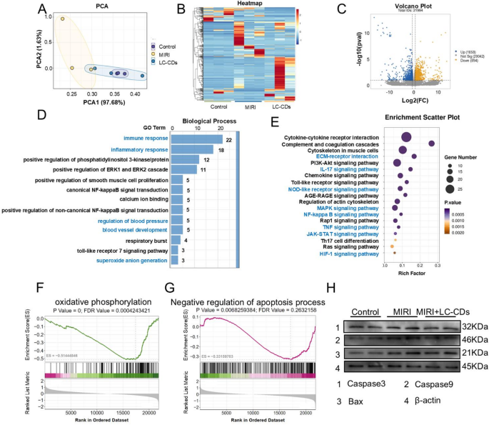
To deeply discover the particular mechanisms underlying the therapy of MIRI by LC-CDs, this examine performed RNA sequencing (RNA-seq) evaluation on the handled rat hearts. The outcomes of principal part evaluation (PCA) clearly introduced the variations among the many three therapy teams: the Management group, the MIRI group, and the LC-CDs therapy group. Amongst them, the MIRI group exhibited essentially the most pronounced variations in gene expression in comparison with the opposite two teams, whereas the guts tissue handled with LC-CDs displayed a profile nearer to the management group (Fig. 8A). The gene-expression heatmap additional revealed that there have been vital gene-expression variations among the many three therapy teams (Fig. 8B). Though there have been additionally sure gene-expression variations among the many three samples inside every group, contemplating that this distinction may be attributed to the pure inter-individual variations, it had no substantial influence on the ultimate outcomes. Subsequently, the gene-expression profiles of rat hearts handled with LC-CDs have been in contrast with these of the MIRI group. The volcano plot revealed a considerable variety of genes with differential expression throughout the 2 pattern teams (Fig. 8C). After therapy with LC-CDs, a complete of 1058 genes have been upregulated, and 894 genes have been downregulated. The outcomes of gene ontology (GO) and Kyoto Encyclopedia of Genes and Genomes (KEGG) enrichment analyses for these differentially expressed genes are introduced as follows. Within the GO evaluation, organic processes have been the important thing focus. The outcomes revealed that differentially expressed genes have been notably related to organic processes together with immune and inflammatory responses, blood strain regulation, blood vessel growth, and superoxide anion manufacturing (Fig. 8D). This means that LC-CDs primarily modulate illness development throughout MIRI restore by concentrating on pathways concerned in immune regulation and superoxide radical manufacturing. Within the KEGG enrichment evaluation, the particular signaling pathways affected by LC-CDs have been recognized, together with ECM-receptor interplay, IL-17 signaling pathway, NOD-like receptor signaling pathway, MAPK signaling pathway, NF-kappa B signaling pathway, TNF signaling pathway, JAK-STAT signaling pathway, and HIF-1 signaling pathway (Fig. 8E). Subsequent, to additional confirm the impact of LC-CDs on the aforementioned signaling pathways, we carried out a extra in-depth statistical evaluation of the important thing downstream genes inside these signaling pathways. The outcomes confirmed that after therapy with LC-CDs, the expression ranges of the important thing genes in these pathways all returned to the identical state as these of the management group. This outcome strongly signifies that LC-CDs can successfully regulate the related signaling pathways (Fig. S7). As well as, gene-set enrichment evaluation (GSEA) offered a complete evaluation of differential gene expression on the gene-set degree. The GSEA outcomes confirmed that two pathways, oxidative phosphorylation and Adverse regulation of the apoptosis course of, underwent vital modifications after LC-CDs therapy (Fig. 8F, G). This discovery aligns with the outcomes of earlier network-pharmacology analyses concerning the mechanisms by which Ligusticum Chuanxiong impacts MIRI. Moreover, western blotting outcomes demonstrated that LC-CDs inhibited apoptosis by means of downregulation of key proteins, together with Caspase3, Caspase9, and Bax (Fig. 8H).
RNA sequencing to discover organic mechanisms of LC-CDs. (A) PCA evaluation of the Management, MIRI, and MIRI + LC-CDs teams. (B) Warmth map of genes in several remedies. (C) Volcano plots of all genes within the MIRI + LC-CDs group and the MIRI group. The blue dots represented the upregulated otherwise expressed genes, the yellow dots have been downregulated otherwise expressed genes, and the gray dots have been non-differentially expressed genes. (D) GO enrichment of differential genes in MIRI + LC-CDs and MIRI teams. (E) KEGG enrichment of differential genes in MIRI + LC-CDs and MIRI teams. (F and G) GSEA enrichment evaluation of differentially expressed genes (oxidative phosphorylation and Adverse regulation of apoptosis course of). (H) Expression of key proteins within the apoptosis pathway (Caspase3, Caspase9, and Bax)
Biosafety analysis of LC-CDs
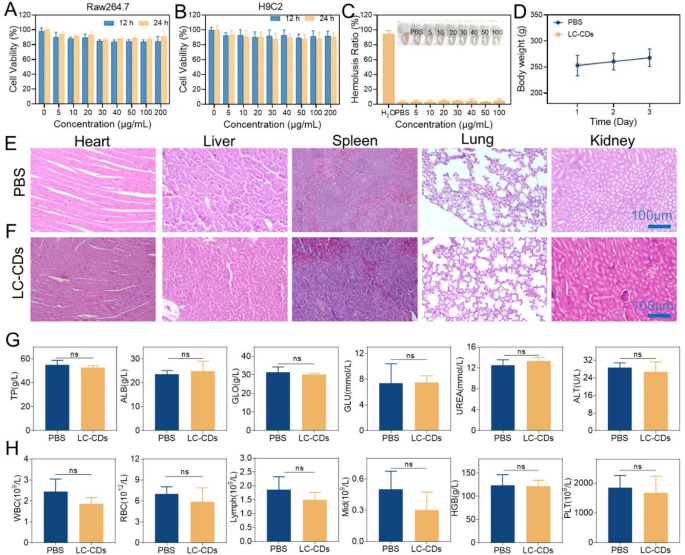
The biosafety of ultrasmall carbon nanoparticles has at all times been a spotlight within the analysis discipline. This examine first investigated the results of LC-CDs on Raw264.7 cells and H9C2 cells on the mobile degree. Encouragingly, even when the focus of LC-CDs reached 200 µg/mL, the MTT assay outcomes confirmed that after 24-hour co-incubation with Raw264.7 cells, the cell viability remained larger than 80% (Fig. 9A). Equally, within the H9C2 cell experiment, H9C2 cells additionally maintained good cell viability after co-incubation with LC-CDs (Fig. 9B). Relating to blood security, it was evaluated by means of a hemolysis experiment. After co-incubating totally different concentrations of LC-CDs with blood cells for 4 h, no irregular phenomena have been noticed within the blood cells, indicating that LC-CDs had good security within the blood (Fig. 9C). To evaluate the in vivo security of LC-CDs in animals, LC-CDs have been injected in situ into the rat hearts. After three consecutive days of commentary, the drug-administered group exhibited a standard progress development in physique weight in comparison with the wholesome group (Fig. 9D). Pathological sections of the primary organs of rats, together with the guts, liver, spleen, lungs, and kidneys, have been ready and noticed by H&E staining. The findings indicated that the guts tissue and different main organs within the drug-administered group exhibited no vital pathological alterations, confirming that LC-CDs didn’t induce extreme injury to those organs (Fig. 9E, F). Moreover, the results of LC-CDs on the capabilities of the primary organs have been evaluated by means of blood biochemical and blood routine assessments. Evaluation of blood biochemical indicators confirmed no vital variations in ranges of Whole Protein, Alanine Aminotransferase, and different markers between the drug-administered group and the management or wholesome teams, suggesting that LC-CDs didn’t considerably have an effect on liver operate (Fig. 9G). The Albumin and Globulin indicators additionally confirmed no variations, suggesting that LC-CDs didn’t set off an immune response in rats. The conventional Glucose and Urea indicators indicated that the blood-glucose degree and kidney operate of the drug-administered group of rats weren’t impaired (Fig. 9G). The blood-routine indicators confirmed that there have been no apparent variations within the blood situations between the LC-CDs-treated group of rats and the wholesome rats (Fig. 9H). In abstract, the above outcomes totally display that LC-CDs possess wonderful biosafety.
Biosafety analysis of LC-CDs. (A) Cell viability of various concentrations of LC-CDs incubated with Raw264.7 for 12 h and 24 h. (B) Cell viability of various concentrations of LC-CDs incubated with H9C2 for 12 h and 24 h. (C) Hemolysis check. (D)Adjustments in physique weight of rats in several therapy teams (n = 3 mice). (E) H&E staining of main organs for biosafety in wholesome teams of mice. (F) H&E staining of main organs for biosafety within the LC-CDs teams of mice. (G) Blood biochemistry evaluation outcomes of the above mice. (H) Full blood panel evaluation outcomes of LC-CDs-treated mice. Statistical comparisons have been made utilizing one-way ANOVA and t-test. “ns” represents “no statistically vital distinction”. Knowledge are expressed as imply ± SD (n = 3)


