Preparation and characterization of CCDs
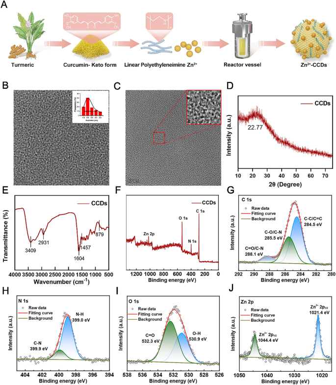
Utilizing curcumin powder as the principle uncooked materials, mixed with zinc ions supplied by zinc acetate and nitrogen wealthy polyethyleneimine, a brownish yellow carbon dots powder was lastly obtained by a traditional one-step hydrothermal methodology (Fig. 1A). Through the synthesis course of, some properties of the uncooked supplies had been properly transferred to the brand new merchandise. On the similar time, numerous parts and electron pairs endure response pairing and binding, giving CCDs new and distinctive properties.
Characterization of CCDs. (A) Schematic illustration of the preparation strategy of CCDs; (B, C) TEM pictures and nanoparticle dimension evaluation; (D) XRD sample of CCDs; (E)FT-IR spectra of CCDs; (F) XPS full-scan spectrim of CCDs; XPS high-resolution scans of C1s (G), N1s (H), O1s (I) and Zn 2p (J) of CCDs.
Fig. 1B and C are TEM pictures of composite nanoparticles, exhibiting that CCDs had been spherical and had a uniform particle dimension distribution, with a mean of two.65 ± 0.3 nm. It may be seen that CCDs had detailed lattice fringes, and the lattice spacing corresponds to the (001) crystal airplane of graphite at 0.21 nm, which was just like the outcomes reported within the literature [23]. The XRD sample of the composite nanoparticles in Fig. 1D confirmed a transparent peak of CCDs at 22.77 °, similar to the (002) crystal airplane of graphite carbon, indicating that CCDs had a whole crystal construction and have been efficiently ready [24]. The floor of CCDs had a optimistic potential of 28.9 (Fig. S1), and the presence of a number of useful teams on the floor facilitates the response of CCDs to laser irradiation in numerous wavelength bands. Research have proven that CCDs with optimistic potential can firmly bind to bacterial surfaces with adverse potential, bettering their bactericidal efficiency [25].
The FT-IR spectrum proven in Fig. 1E exhibited peaks at 3409, 3255, 2931, 1604, and 1457 cm− 1, similar to the stretching vibrations of – OH/- NH, C-H, C = C, and N-H teams, respectively. The height at 879 cm− 1 may very well be thought-about a attribute peak of ZnO, which is said to particular vibration modes within the ZnO crystal construction. Subsequently, the numerous water solubility of CCDs may be attributed to the presence of plentiful hydrophilic teams on their surfaces, which additionally confirmed the existence of Zn [26]. The chemical composition and useful teams of CCDs had been analyzed utilizing X-ray photoelectron spectroscopy (XPS). The XPS full spectrum in Fig. 1F confirmed that CCDs are primarily composed of C, N, O, and Zn parts. The high-resolution XPS spectrum of C1s may be decomposed into three peaks with binding energies of 288.1 eV, 255.5 eV, and 284.5 eV, respectively. These peaks represented C = O/C-N, C-O/C-N, and C-C/C = C, respectively (Fig. 1G). The N1s band deconvolution peaks at 399.9 eV and 399.0 eV correspond to graphite nitrogen and pyrrole nitrogen, respectively (Fig. 1H) [27]. The peaks of O1s at 532.3 eV and 530.9 eV correspond to C = O and O-H (Fig. 1I) [26]. Fig. 1J confirmed the high-resolution XPS spectrum of Zn2p, with two peaks at binding energies of 1044.4 eV and 1021.4 eV. These peaks corresponded to the spin orbit dipoles of Zn 2p3/2 and Zn 2p1/2, respectively, indicating that zinc doped CCDs include zinc within the two optimistic valence state [28]. The plentiful nitrogen in CCDs comes from polyethyleneimine, which might present the mandatory parts for the synthesis of carbon dots. In the meantime, nitrogen atom doping, as an environment friendly and handy methodology, supplies plentiful energetic websites and useful teams on the floor of CCDs, which broadens the appliance scope of carbon dot nanomaterials. As a result of the doping of nitrogen performs an vital position in bettering the fluorescence properties and photosensitizing exercise of carbon dots, it might result in a 20 nm redshift of the utmost fluorescence emission place of the carbon dots and a rise within the emission depth of the carbon dots, a property that’s of nice significance for using carbon dots as photosensitizers [29]. All these outcomes demonstrated the profitable synthesis of Zn-CCDs nanoparticles.
Antibacterial property
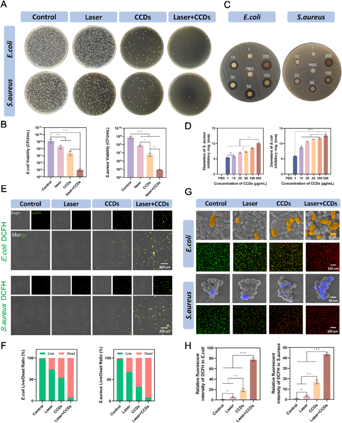
As a result of broken barrier, exposing the wound to the advanced each day setting will increase the danger of bacterial an infection. S. aureus is ubiquitous on the floor of the pores and skin and within the dwelling setting and is among the most typical infectious brokers of the pores and skin [30, 31]. As well as, many research have proven that E. coli is growing the incidence of wound infections [32, 33]. Subsequently, S. aureus and E. coli had been chosen as consultant strains of Gram-positive and Gram-negative micro organism for the examine of the antimicrobial properties of CCDs in PDT of contaminated wounds. The CCDs composites with an general positively charged nature had been firmly connected to the negatively charged bacterial surfaces by electrostatic interplay. On the similar time, part of the micro organism ingested the nanoscale CCDs into the intracellular area, and PDT impact was produced beneath laser irradiation to realize the killing impact. As proven in Fig. 2C, totally different concentrations of CCDs fashioned inhibitory rings with various diameters, indicating that the inhibitory impact of CCDs on micro organism was stronger because the focus elevated (1–200 µg/ml). Digital pictures of S. aureus and E. coli forming colonies on agar plates in several therapy teams are proven in Fig. 2A. The variety of colonies within the Laser + CCDs group was considerably lower than that of the management group, with a discount of three–4 orders of magnitude. It may be seen that laser irradiation or CCDs alone had some inhibitory impact on the expansion of each micro organism, however the impact was poor.
Antimicrobial properties. (A) Plate rely pictures of E. coli and S. aureus in every group; (B) Quantitative evaluation of the variety of E. coli and S. aureus colonies; (C) Ring of inhibition pictures of E. coli and S. aureus in every group. The totally different situations are PBS buffer or CCDs answer with concentrations of 10, 20, 50, 100, 200 µg/mL; (D) Quantitative evaluation of diameters of inhibition ring of E. coli and S. aureus with totally different focus of CCDs; (E) ROS probe fluorescent staining pictures of E. coli and S. aureus in every group; (F) Quantitative ratios of dwell and lifeless E. coli and S. aureus; (G) SEM pictures and dwell/lifeless fluorescent staining pictures of E. coli and S. aureus in every group; (H) Quantitative evaluation of the depth of ROS fluorescence of E. coli and S. aureus. (*p < 0.05, **p < 0.01, ***p < 0.001)
CCDs, as a novel photosensitizer, are able to producing ROS beneath laser irradiation for photodynamic results. As a way to examine the particular mechanism of bacterial killing by Laser + CCDs, we used DCFH-DA as a reactive oxygen probe to probe the ROS manufacturing in micro organism after totally different remedies. The presence of ROS in micro organism oxidizes the non-fluorescent DCFH-DA to the robust inexperienced fluorescent substance DCF [34]. Fig. 2E confirmed a weak inexperienced fluorescence in E. coli and S. aureus after ingestion of CCDs, and a shiny inexperienced fluorescence in nearly all micro organism after PDT, representing the manufacturing of a considerable amount of ROS after photodynamic remedy. Fig. 2G confirmed the SEM pictures of the 2 micro organism after PDT, with pseudo-colors getting used to focus on the areas of curiosity. It may be seen that the floor of the micro organism within the Management group was clean and full, with clear and intact boundaries, in a position to kind a whole biofilm construction. Within the Laser + CCDs group, it was apparent to see that the bacterial cell membranes had been wrinkled, collapsed, with blurred boundaries, and in some circumstances the contents had been within the type of a jet, which is a typical manifestation of the extreme harm to the micro organism attributable to the excessive focus of ROS. The floor of S. aureus was twisted and compressed and deformed, however mainly remained intact and spherical. In distinction, the diploma of destruction of E. coli was extra vital, the cell membrane was strongly contracted and ruptured, and the shell misplaced its intact morphology. This distinction could also be on account of the truth that the phosphomimic acid cell wall of the outer layer of Gram-positive micro organism restricted the enlargement and rupture of the bacterial physique to a sure extent. Part of the massive quantity of ROS generated by photodynamic pressure acted straight on the cell membrane construction, whereas the opposite half attacked the organelles and nucleic acids, inflicting irreversible harm and thus killing the micro organism. That is the traditional sort I mechanism in PDT, which performed an vital position in the entire therapy system [35]. This therapeutic mechanism considerably slowed the event of bacterial drug resistance and prevents the formation of bacterial biofilms. We hypothesize that Zn2+ was launched from the composite nanoparticles and disrupted the cell membrane by neutralizing the adverse cost of the cell membrane [36]. On the similar time, the composition of CUR may inhibit the adhesion of micro organism to a sure extent, significantly decreasing the danger of bacterial an infection [37].
In Fig. 2G, dwell/lifeless fluorescence staining was carried out on the micro organism in several therapy teams to visually distinguish the survival standing of micro organism by crimson and inexperienced fluorescence. In contrast with the Management group and the Laser group, the remainder of the therapy teams had apparent crimson fluorescence, indicating that the inactivated micro organism accounted for a terrific proportion. In distinction, there was nearly no inexperienced fluorescence within the Laser + CCDs therapy group, and this killing confirmed the identical pattern within the quantitative evaluation of Fig. 2F. Some research have already reported that PDT has totally different selectivity for Gram-negative and Gram-positive micro organism on account of variations within the floor construction of bacterial cell membranes. Normally, photosensitizers usually tend to penetrate the porous cell partitions of Gram-positive micro organism to provide ROS to behave as bactericidal brokers [38, 39].
Exploration of the mechanism of CCDs-mediated PDT additionally revealed that the ROS produced by this therapy modality inhibits the formation of bacterial biofilm, which improves the permeability of CCDs and enhances the killing impact on deep-seated micro organism [40]. On the similar time, this therapy reduces the danger of creating drug resistance by not focusing on the attribute levels of the bacterial metabolic part and impeding the formation of biofilm——the barrier. The outcomes of biofilm formation inhibition experiments confirmed (Fig. S2) that the density and construction of the 2 bacterial biofilms had been considerably disrupted after Laser + CCDs therapy and antibiotic Ciprofloxacin therapy, exhibiting nearly the identical pattern, indicating that the impact of CCDs-mediated PDT is nearly corresponding to that of the usual antibiotics by way of antimicrobial exercise. Subsequently, CCDs as photosensitizers can successfully induce ROS burst to inhibit biofilm formation and considerably kill micro organism.
Fig. S3 confirmed the outcomes of circulation cytometry evaluation of CCDs mediated PDT on E. coli and S. aureus. It may be seen that the majority (99.1%, 99.5%) of the micro organism within the management group didn’t endure apoptosis, which implies that a lot of the micro organism within the group had been in a viable state. Each the Laser group (3.34%, 8.17%) and the CCDs group (19.5%, 20.4%) confirmed various numbers of stained cells with apoptotic alerts, however apoptotic alerts had been stronger within the CCDs group, which suggests each of the remedies can each kill sure micro organism, however the killing capability of CCDs is stronger. After the addition of laser irradiation, the survival charge of each micro organism within the Laser + CCDs group decreased dramatically, exhibiting comparable outcomes to the antibiotic Ciprofloxacin. Amongst them, the apoptotic cells of E. coli elevated to 32.3% (37.8% within the Ciprofloxacin group) whereas these of S. aureus elevated to 31.4% (36.9% within the Ciprofloxacin group). The outcomes confirmed that CCDs-mediated PDT had glorious antibacterial results, even just like these of ordinary antibiotics, and was efficient in inducing apoptosis and inhibiting additional propagation of micro organism. CCDs adhered to the bacterial organisms and entered into the intracellular compartment, after which ROS generated by the PDT broken the interior biomolecules of micro organism and finally led to bacterial loss of life, which is in step with the conclusions drawn from the earlier research [41].
Promote cell migration and vascularization
Broken tissues start to proliferate and restore after the inflammatory part, and blood vessels are vital channels for transporting vitamins and development elements [42]. Classical scratch and transwell migration assays had been chosen to look at the angiogenic potential of HUVEC and L929 cells after PDT, and a matrigel-based tube formation assay for HUVEC was designed to additional examine.
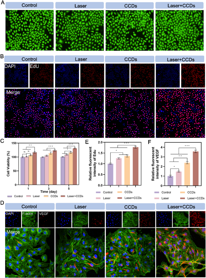
The cytotoxicity of CCDs was first explored by customary CCK-8 assay. Fig. S4 confirmed that CCDs at totally different concentrations didn’t exhibit vital toxicity after 1, 3 and 5 days of co-culture with HUVEC. Apparently, CCDs at a focus of 200 µg/ml had been in a position to considerably promote the proliferation of endothelial cells to some extent. This focus was chosen as the usual focus for subsequent assays. Fig. 3C and Fig. S5 confirmed that the Laser + CCDs therapy group promoted the proliferation of HUVEC and L929 cells, respectively. The CCDs therapy group additionally barely raised the viability of each cells. The remaining therapy teams confirmed no vital distinction. Based mostly on the outcomes of dwell/lifeless cell staining of HUVEC cultured with CCDs for twenty-four h, nearly no lifeless cells (crimson fluorescence) had been noticed in every therapy group, as proven in Fig. 3A. EdU staining of HUVEC confirmed comparable outcomes to these of CCK-8. EdU is a thymine deoxyribonucleoside analog that’s able to changing the thymine deoxy nucleus doped into de novo and synthesized DNA throughout DNA synthesis. Subsequently, the quantity of newly synthesized DNA was examined by the EdU-related package to replicate the proliferation standing of the cells. The outcomes confirmed (Fig. 3B) that the Laser + CCDs group exhibited shiny crimson fluorescence, and the remainder of the therapy teams had weaker fluorescence depth (Fig. 3E), indicating that CCDs within the presence of laser irradiation might certainly promote HUVEC propagation and facilitate wound restoration.
Promotion of HUVEC proliferation and angiogenic protein expression. (A) Dwell/lifeless staining pictures of HUVEC; (B) Fluorescence pictures of HUVEC EdU staining in every group; (C) Cell proliferation in CCK8 assay at 1, 3 and 5 days; (D) Immunofluorescence staining pictures of VEGF protein in every group; (E) Quantitative evaluation of fluorescence depth of EdU staining in every group; (F) Quantitative evaluation of immunofluorescence depth of VEGF protein in every group. (*p < 0.05, **p < 0.01, ***p < 0.001)
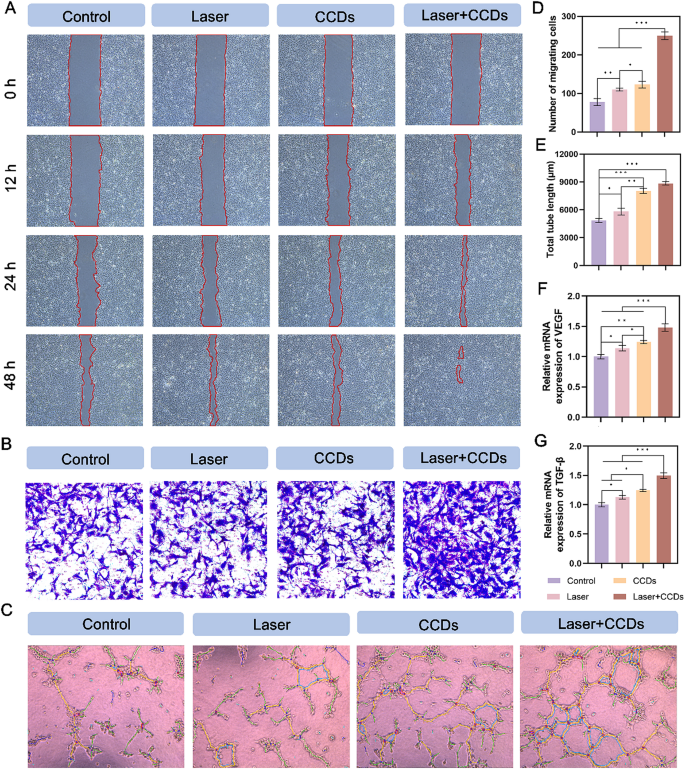
Scratch therapeutic by endothelial cells is central to the method of tissue re-formation throughout wound therapeutic. As proven in Fig. 4A and Fig. S6A, each HUVEC and L929 cells within the Laser + CCDs group exhibited optimum therapeutic after therapy. Notably, the CCDs group additionally confirmed some promotion within the scratch assay for each cells. The info in Fig. S5 and Fig. S6D quantified the therapeutic charge of scratches, exhibiting the identical pattern. After 24 h of incubation, the migration charges of HUVEC and L929 cells had been considerably larger within the Laser + CCDs group than within the different therapy teams (Fig. 4B and S6B). Endothelial cells are elements of blood vessels and play an important position in angiogenesis. The matrix gel tube formation assay carried out to evaluate the angiogenic capability confirmed (Fig. 4C) that Laser + CCDs-treated HUVEC confirmed a big improve in tube size and variety of junctions after 6 h in contrast with the Management group (Fig. 4E and S8), which can be attributed to the discharge of Zn2+ in addition to the era of managed ROS. Total, CCDs have the flexibility to advertise cell migration beneath laser irradiation, which is helpful for accelerating the wound therapeutic course of.
Promotion of HUVEC migration and angiogenic property. (A) Consultant pictures of HUVEC scratching assay, the crimson underlined portion represents the scratched space; (B) Consultant pictures of HUVEC transwell migration assay in every group; (C) Photographs of HUVEC tube formation assay; (D) Quantitative evaluation of the variety of migrating cells in every group; (E) Quantitative evaluation of the full tube size formation in every group; (F) Quantitative evaluation of relative mRNA expression of VEGF; (G) Quantitative evaluation of relative mRNA expression of TGF-β. (*p < 0.05, **p < 0.01, ***p < 0.001)
As a widely known angiogenesis associated issue, vascular endothelial development issue (VEGF) induces proliferation, migration, and angiogenesis of epidermal and endothelial cells. Throughout angiogenesis, VEGF is important for the recruitment, upkeep, proliferation, migration and differentiation of endothelial cells [43, 44]. Subsequently, we expressed the expression of VEGF protein in cells of various therapy teams by fluorescence staining (Fig. 3D and S6C). The outcomes confirmed that HUVEC and L929 cells exhibited shiny crimson fluorescence within the cytoplasm after PDT, and CCDs-treated group additionally had robust crimson fluorescence, whereas those within the Management group confirmed nearly no expression. The quantitative evaluation of fluorescence depth in Fig. 3F and Fig. S6F is per the above. The relative gene expression ranges of VEGF and TGF-β within the two forms of cells (Fig. 4F and G and S9, S10) had been nearer within the CCDs and Laser + CCDs teams, and considerably larger than these within the Management and Laser teams. Zinc, as a necessary hint aspect, performs an vital organic perform within the upkeep of the venous vascular community and the event of hematopoiesis [45]. Our examine confirmed that CCDs stimulate angiogenesis by slowly releasing Zn2+, which is per earlier studies [46]. And the aforementioned toxicity take a look at additionally confirmed that Zn2+ within the composite nanoparticles additionally didn’t have an effect on the conventional cell viability. The above outcomes indicated that CCDs exhibit advanced and energetic regulation of angiogenesis after laser irradiation.
The potential mechanism of accelerated contaminated wound therapeutic
Wound therapeutic is a posh course of involving interactions between totally different cell sorts, development hormones, cytokines, antioxidants, and a gentle provide of steel ions (e.g., calcium, zinc, and magnesium). After pores and skin harm, a variety of mobile programs and signaling pathways are activated within the wound to guard the physique [47]. As a way to elucidate the doable regulatory mechanisms of Zn2+-doped curcumin carbon dots to advertise contaminated wound therapeutic, we carried out transcriptome evaluation on cells from two totally different therapy teams (Management and Laser + CCDs teams).
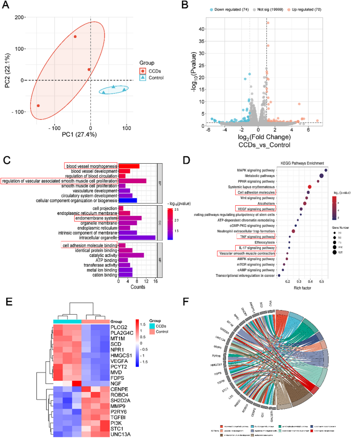
Principal Part Evaluation (PCA) outcomes confirmed that samples from every group had been clustered independently, verifying that the 2 therapy teams did produce variations (Fig. 5A). Volcano plots exhibiting up- and down-regulated genes generated after totally different remedies within the Management and Laser + CCDs teams exhibited vital differential gene expression between the totally different teams (Fig. 5B). To additional analyze these differential gene expressions, we carried out Gene Ontology (GO) enrichment evaluation. GO sometimes characterizes genes at three ranges: mobile element (CC), organic course of (BP), and molecular perform (MF). Fig. 5C confirmed the highest enriched gadgets in every class, which primarily embrace down-regulated expression of immune response, inflammatory response elements and up-regulated vascular morphology and proliferation of vascular clean muscle, endothelial phylogeny and cell adhesion.
Gene expression profiles and regulatory mechanisms in selling infectious wound therapeutic. (A) PCA evaluation of samples; (B) Volcano plot with up- and down-regulated genes; (C) Consultant high 22 differentially expressed phrases analyzed by the Gene Ontology (GO) enrichment methodology; (D) Consultant high 20 up- or down-regulated pathways analyzed by KEGG pathway methodology; (E) The Heatmap evaluation and (F) circus of differentially expressed genes concerned in a number of pathways
In the meantime, Kyoto Encyclopedia of Genes and Genomes (KEGG) enrichment evaluation was utilized to investigate the potential signaling pathways. Fig. 5D confirmed intimately the pathways with excessive relevance and their names, and the up-regulated signaling pathways embrace VEGF signaling pathway, cell adhesion molecule signaling pathway and mitogen-activated protein kinase (MAPK) signaling pathway.
It’s well-known that the VEGF signaling pathway is carefully associated to collagen deposition and angiogenesis. And the MAPK signaling pathway promotes keratinocyte proliferation and migration, and performs an vital position in transmitting extracellular stimulatory alerts to cells and mediating mobile organic responses (e.g., cell development, migration, proliferation, differentiation, and apoptosis). However, CCDs photodynamic remedy additionally down-regulated among the pathways, together with the IL-17 signaling pathway, and the tumor necrosis issue (TNF) signaling pathway, that are related to the inflammatory response and regulation of tissue destruction. The heatmap in Fig. 5E confirmed the names of the genes with particular adjustments for main variations within the above pathways. Particular names and interconnections concerning the genes screened for larger differential expression by GO enrichment evaluation are displayed in Fig. 5F. These outcomes instructed that the VEGF signaling pathway could also be activated throughout PDT with CCDs to advertise mobile angiogenesis, thereby accelerating wound therapeutic.
MAPK is essential in transmitting alerts of extracellular stimuli to the cells and mediating mobile organic responses (e.g., cell development, migration, proliferation, differentiation and apoptosis). Mammalian MAPKs may be categorized into 4 subfamilies: ERK1/2, p38, JNKs and ERK5 [48]. A number of differential genes of the ERK pathway had been enriched within the MAPK signaling pathway. VEGF is an important angiogenic stimulator and performs vital roles in angiogenesis and neointima formation, together with inflicting cell proliferation, inhibiting apoptosis, growing vascular permeability, and vasodilation. Genes upregulated within the VEGF/VEGFR2 pathway embrace VEGFA, PLCG2, and PLA2G4C. Amongst them, PLA2G4C can be concerned within the ERK pathway.
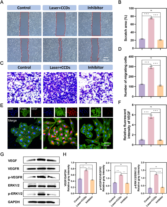
To analyze whether or not the MAPK and VEGF signaling pathways had been potential mechanisms and additional validate the above speculation to advertise infectious wound therapeutic, we validated them by way of genes and proteins in addition to phenotypes. VEGFR2 inhibitor (SU19498) was used to check the reliability of the sequencing outcomes. The scratch take a look at outcomes after 12 h in Fig. 6A confirmed that the scratch space of the Management group and the Inhibitor group has not modified a lot in comparison with 0 h, with solely a hint quantity of therapeutic. The Laser + CDs group confirmed a big selling impact on therapeutic. The migration assay in Fig. 6C additionally confirmed the identical pattern, with solely the Laser + CCDs group exhibiting numerous cells migrating to the decrease chamber. This proves that VEGFR2 inhibitor hindered the signaling course of on this pathway, thereby inhibiting the expansion and migration of HUVEC cells. The above results had been additionally demonstrated by VEGF immunofluorescence and immunoblotting (Fig. 6E and G), confirming the significance of VEGF in endothelial cell development and numerous physiological actions. The VEGF/VEFGR2 pathway is carefully associated to the MEK1/2/ERK1/2 signaling pathway, and one of many downstream pathways of the VEGF signaling pathway is to mediate cell proliferation by the ERK pathway [49]. By WB detection of VEGF, VRGFR2, phosphorylated VRGFR2, phosphorylated ERK1/2, and ERK1/2, we discovered that the protein expression ranges of the Management group and pathway inhibitor group had been nearly the identical, whereas the expression of every index was considerably elevated within the PDT group with CCDs. It’s significantly noteworthy that the appliance of VEGFR2 inhibitor (SU19498) can successfully inhibit the phosphorylation of VEGFR2, leading to a big lower within the expression ranges of downstream VEGF proteins p-VEGFR2/VEGFR2 and p-ERK1/2/ERK1/2 within the Inhibitor group. In abstract, the above outcomes indicated that CCDs promote endothelial cell development and wound therapeutic by the VEGF/VEGFR2/ERK1/2 pathway.
Verification of VEGF signaling pathway mechanism in HUVEC. (A) Consultant pictures of scratching assay, the crimson underlined portion represents the scratched space; (B) Quantitative evaluation of scratch space closure in every group; (C) Consultant pictures of migration assay in every group; (D) Quantitative evaluation of the variety of migrating cells in every group; (E) Immunofluorescence staining pictures of VEGF protein in every group; (F) Quantitative evaluation of immunofluorescence depth of VEGF protein in every group;(G) Expression of VEGF, VEGFR, ERK1/2, phospho-VEGFR, -ERK1/2 and GAPDH, in addition to the grey depth evaluation in HUVEC; (H) Quantitative evaluation of (G). (*p < 0.05, **p < 0.01, ***p < 0.001)
The outcomes instructed that CCDs photodynamic remedy exerts its position in selling vascular endothelial cell development by modulating VEGF/VEGFR2 and its downstream MEK1/2/ERK1/2 signaling axis. Thus, wound therapeutic can’t be achieved with out exact regulation together with hemostasis, irritation, proliferation and reworking sequences.
Analysis of in vivo contaminated wound therapeutic
Based mostly on the efficacy of CCDs with PDT in killing micro organism and selling the proliferation and restore of endothelial cells obtained in vitro, we additional used contaminated wound rats to check their restore impact in vivo.
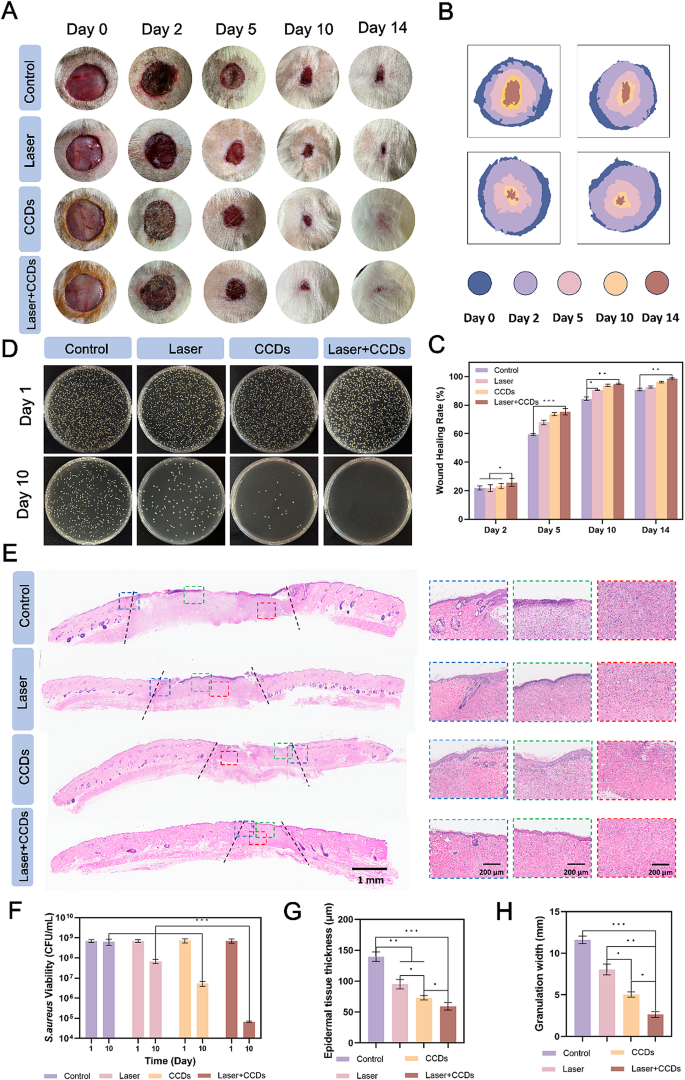
First, a whole-layer rat pores and skin defect mannequin with a diameter of two cm was created, and S. aureus bacterial suspension was utilized to the wound floor to determine an an infection mannequin. Saline and CCDs had been injected into the contaminated wounds for therapy (named Management group, Laser group, CCDs group, and Laser + CCDs group, respectively), and the parameters of the laser used within the therapy had been 660 nm, 10 min, and 500 mW/cm2. Digital pictures of the wound space at totally different time factors and overlapping pictures of the simulated therapeutic are proven clearly in Fig. 7A and B. After 24 h, the wound the wound grew to become visibly contaminated. The outcomes confirmed that on the finish of the primary therapy (day 2), the floor of the Management and Laser teams was ulcerated and oozing, bleeding was obvious, and no apparent scab formation was seen, whereas the CCDs and Laser + CCDs teams confirmed apparent indicators of therapeutic, with the bleeding stopping and the formation of thicker therapeutic scabs. After 14 days, the injuries of the CCDs and Laser + CCDs teams had been nearly healed however accompanied by various levels of scar formation. In distinction, wounds within the Management and Laser teams remained open, and the newly fashioned tissue was considerably thinner and redder. The proportion of wounds healed in every group of rats is quantified in Fig. 7C. Important variations had been noticed between teams after 2 days, with the Management group having the slowest therapeutic charge values in any respect time factors. 92.65%, 96.05%, and 98.7% of wounds had been healed on the final time level within the Laser group, the CCDs group, and the Laser + CCDs group, respectively. To straight characterize the an infection, tissues from the wound space had been homogenized and incubated on agar plates. Fig. 4D and F confirmed the colony diagrams and quantitative outcomes of localized micro organism in all wounds after day 1 and day 10, respectively. The outcomes confirmed that numerous colonies with floor bacterial colonization and development had been noticed in all therapy teams at day 1. Whereas, on day 10, the variety of colonies within the Laser + CCDs group was considerably lowered, and quantification confirmed that S. aureus was lowered by nearly 4 orders of magnitude. The above outcomes indicated that PDT with CCDs has the potential to regulate wound an infection and help therapeutic.
Contaminated wounds therapeutic in vivo. (A) Consultant digital pictures of every group of wounds at day 0, 2, 5, 10, and 14; (B) Schematic illustration of overlapping wound areas at consultant time factors; (C) Quantification of the closed wound space share; (D) Plate rely pictures of S. aureus of wounds in every group at day 1 and 10; (E) Consultant pictures of the H&E staining in every group, exhibiting pores and skin tissue, the general construction of the neoplastic epithelium on the wound edge (within the blue dashed field), the epithelium within the middle of the wound (within the crimson dashed field), and the subepithelial layer (within the inexperienced dashed field); (F) Quantitative evaluation of the variety of S. aureus colonies in every group; (G) Quantitative evaluation of the new child epithelial tissue thickness and (H) the granulation tissue width in every group. (*p < 0.05, **p < 0.01, ***p < 0.001)
As well as, to additional discover the advanced organic processes together with irritation, proliferation, and reworking in the course of the therapeutic of contaminated wounds handled with CCDs.
in PDT, we evaluated the histological adjustments after 14 days. At day 14 post-treatment, hematoxylin-eosin (H&E) staining of the wound and surrounding space confirmed histologically related wound therapeutic, corresponding to epithelial re-formation and granulation tissue, as proven in Fig. 7E. The outcomes of the H&E staining of the wound and surrounding space confirmed that the wound therapeutic course of was not so simple as that of the Management group. The granulation tissue width (distance between the black dotted traces) was considerably lowered within the CCDs group and narrowest within the Laser + CCDs group in comparison with the Management and Laser teams, with quantitative outcomes exhibiting the identical pattern (Fig. 7H). The blue dashed field in Fig. 7E confirmed the conventional epithelium versus the broken epithelium, and it may be seen that the photodynamically handled tissues appeared as clean and flat epithelium with neoplastic hair follicles, which is nearer to the state of regular pores and skin. In distinction, the opposite therapy teams had disorganized cells within the basal layer, disorganized collagen, and clear demarcation from the conventional epithelium. The crimson dashed field illustrated the discount of inflammatory cell infiltration and the looks of extra fibroblast migration within the epithelium within the Laser + CCDs group after photodynamic therapy. The inexperienced dashed field confirmed that the management group had edema, discontinuity and dense inflammatory cell bands within the epithelium. The Laser + CCDs group had a thinner epithelium with no seen inflammatory cells and extra mature tissue reworking. Quantitative evaluation of epithelial tissue thickness was proven in Fig. 7G. Thicker epithelial tissue thickness was noticed within the Management group (138 ± 12 μm) and Laser group (94 ± 7 μm).
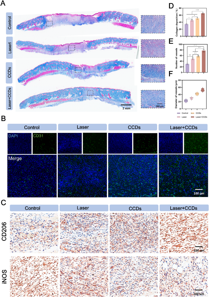
Collagen deposition is a key determinant of pores and skin power and maturity [50]. Masson staining in Fig. 8A gave a extra particular image of the quantity of collagen within the new child pores and skin tissue and the association of its fibers. The collagen fibers within the CCDs group and the Laser + CCDs therapy group had been extra dense, well-arranged, and the tissue was extra mature. Quantitative evaluation of collagen deposition in Fig. 8D confirmed the identical outcomes. The foremost collagens within the pores and skin are collagen sorts I and III, which make up about 95% of the pores and skin’s collagen composition. These two forms of collagens play an vital position in sustaining the elasticity, integrity and pliability of the pores and skin. Not solely that, however these two most vital forms of collagens are additionally beneath dynamic change in the course of the wound therapeutic course of along with the rise within the general quantity of collagen [51]. Subsequently, Sirius crimson staining was utilized to differentially stain primarily sort I (crimson/yellow) and sort III (inexperienced) collagens primarily based on the totally different traits of collagen polymerization winding and helical association. The outcomes confirmed (Fig. S11) that at day 7, a decrease proportion of sort I and sort III collagen was noticed within the Laser + CCDs group, with an general green-dominated fluorescent sign, which was on account of the truth that sort III collagen was extra predominant within the early wound therapeutic stage. Within the strategy of wound reworking and tissue maturation, sort III collagen was progressively changed by mature sort I. The Laser + CCDs group confirmed an analogous proportion of sort I and sort III collagen as regular pores and skin on day 14, and the general fluorescence sign was reddish-yellow, which indicated that the wound therapeutic after PDT tended to be normalized throughout reconstruction and reworking, and there was no delayed and disordered collagen deposition.
(A) Consultant pictures of Masson staining; (B) Immunofluorescence staining pictures of CD31 expression in every group; (C) Consultant immunohistochemical staining pictures of CD206 and iNOS expression in every group; (D) Quantitative evaluation of collagen deposition in every group; (E) Quantitative evaluation of blood vessel quantity and (F) diameter in every group. (*p < 0.05, **p < 0.01, ***p < 0.001)
The blood vessels within the new tissue can ship important vitamins, development elements and oxygen to the wound web site, and CD31 is a typical marker for vascular endothelial cells [52]. As proven in Fig. 8B, the immunofluorescence pictures confirmed that the Management and Laser teams confirmed few inexperienced fluorescent alerts, whereas the CCDs and Laser + CCDs therapy teams confirmed extra alerts and extra full inexperienced fluorescent rings. Subsequently, we analyzed the quantity and diameter of blood vessels (Fig. 8E and F) and located that may CCDs group and Laser + CCDs therapy group promoted the formation of vascular system. Zn2+ within the composite nanoparticles induced proliferation, migration and angiogenesis of epidermal and endothelial cells [53]. The inflammatory response interval is a stage in regular wound therapeutic, and we explored the inflammatory adjustments throughout wound therapeutic by evaluating two typical inflammation-related elements. In Fig. 8C, the anti-inflammatory issue CD206 was proven to be barely expressed within the management group, whereas the expression confirmed an growing pattern within the Laser, CCDs and Laser + CCDs therapy teams, and the pro-inflammatory issue iNOS confirmed a totally reverse expression within the 4 therapy teams. The indication instructed that PDT successfully elevated the anti-inflammatory degree in vivo and considerably lowered irritation. The above outcomes instructed that CCDs promote therapeutic of contaminated wounds by accelerating the blood provide for neovascularization in addition to reducing the extent of irritation by photodynamic remedy. On the finish of the animal experiments, H&E staining of main organs was carried out (Fig. S14). There have been no variations between all therapy teams, suggesting that CCDs had been non-toxic and properly biologically protected in vivo and might considerably promote contaminated wound therapeutic in a protected method.


